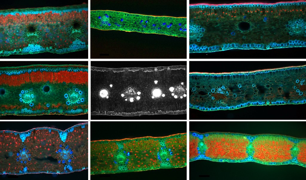

I am quite happy to say that our work on the leaflet anatomy of the Zamiaceae has made the front cover of this issue (volume 181 issue 7) of the International Journal of Plant Sciences. The cover is an artistic rendition of the sections of eight out of nine genera of the Zamiaceae imaged using fluorescence microscopy.
The fresh sections of leaflets of the Zamiaceae present a strong autofluorescence, which allowed us to obtain good details of the anatomical structures (as well as generating beautiful images). These allowed us to show that the anatomy of the leaflets presents a evolutionary signal in the Zamiaceae, supporting some of the results of molecular phylogenetics. These include the grouping of Stangeria with Zamia and Microcycas, the isolated position of Dioon and Bowenia, the close relationship of Encephalartos and Lepidozamia and more.
I wish to thank all the coauthors on this paper for their help and effort, especially the two students Nicola Jelmini and Hanna Neuenschwander who did an excellent job with sectioning and imaging cycad leaflets!

Leave a comment